Electroencephalography (EEG) is the measurement of electrical impulses in the brain. The electrical activity of neurons in the brain produces a current that can be measured by non-invasive electrodes attached to the surface of the scalp. EEG signals reflect neural activity with millisecond temporal resolution, providing a direct non-invasive neural correlate.
In order to record these electrical signals, participants must wear an electrode cap with conductive gel inserted between the cap and scalp, as pictured below. This gel is a saline solution that easily washes off skin and out of hair – there is a sink in the lab specifically for this. In our lab, we use QuickAmp (Brain Products) 64 channel system and BrainVision to analyse the data.
In the Visual Neuroscience lab, we use event-related potentials (ERPs) and visual-evoked potentials (VEPs) to analyse the participant’s response to visual stimuli. EEG is non-invasive, safe and provides an immediate direct response to brain activation.
Psychophysics measures the relationship between physical stimuli and the psychological responses generated. This involves systematic and precise research, computational modelling and mathematical tools. Visual psychophysics, the quantification of visual perception, explores the complex interactions between the eye and the brain.
Psychophysics is an essential discipline for probing perception. Psychophysics places the emphasis on the physical properties of the stimulus and asks participants simple questions, for example determining the point of detection of a stimulus; stimulus recognition; stimulus discrimination or how much sensory information is present.
This field has many practical applications to visual tasks such as driving, reading and visual search. Psychophysical experiments often take place using computers or purpose-built equipment, all of which vary significantly depending on specific project requirements.
What we do:
Functional Near Infrared Spectroscopy (fNIRS): First lab in Scotland to use this technique (set up in 2007). Using this technique we study blood flow in normal and ageing visual brains using mostly standard stimuli for acuity (checkerboards), stereopsis (red/green anaglyphs) and motion coherence (random dot kinematograms).
Event Related Potentials (ERPs) are done to correlate with blood flow in primary visual cortex. We have just acquired a small system to record electroretinograms (ERG). These will also be used to correlate both electrical and metabolic activity in the retina and brain.
Clinically Applicable Studies: Amblyopia and perceptual learning, glaucoma and brain oxygenation, diabetes, retinal perfusion using OCT and OCTA and cerebral oxygenation. Correlation of several of these with psychophysical paradigms.
Optical Coherence Tomography (OCT): The latest arm of our research is to correlate blood flow in the retina using OCT Angiography of the optic disc and macular area with blood flow in the brain using fNIRS. We will correlate ERG activity with blood flow in the retina.
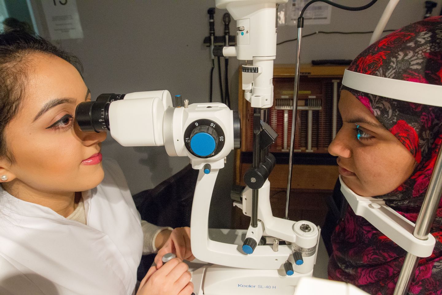
Visual Psychophysics: Using psychophysics we find thresholds in studies of global motion coherence, contrast sensitivity, stereopsis & face perception across the lifespan.
Techniques we use:
Functional Near Infrared Spectroscopy (fNIRS) measures brain activity noninvasively and is unaffected by noise from electromagnetic fields. NIR light penetrates up to 2-3 cm below the surface of the scalp estimating changes in blood oxygenation and photon propagation within the head shows that only haemoglobin and its chromophore concentrations change as a function of brain activity. It equates well with fMRI.
Event Related Potentials (ERPs) are the averaged electrical activity of the brain over time in response to a specific stimulus. They are measured using scalp electrodes.
Electroretinography/electroretinograms (ERG): This technique records the electrical activity of the neural layers of the retina; these layers are the light detecting part of the retina. Their activity helps us see and the ERG detects the electrophysiological function of the retina.
Optical Coherence Tomography (OCT): Images the structure & thickness of the retina non-invasively using eye-safe near-infrared light. OCT angiography uses the variation in OCT signal caused by moving particles, such as red blood cells, as the contrast mechanism for imaging blood flow in the retina.
Visual Psychophysics: Adaptive staircase procedures (or the classical method of adjustment) to measure thresholds to a variety of visual stimuli.
Publications 2010-2020:
- 2020 Aitchison, R.T., Kennedy, G.J., Shu, X. Mansfield DC & Shahani U Sub-clinical thickening of the fovea in diabetes and its relationship to glycaemic control: a study using swept-source optical coherence tomography. Graefes Arch Clin Exp Ophthalmol (2020). https://doi.org/10.1007/s00417-020-04914-2
- 2018 Ward L, Aitchison RT, Hill, G, Imrie, J, Simmers AJ, Morrison G, Mansfield DC, Kennedy G & Shahani U Effects of Glaucoma and Snoring on Cerebral Oxygenation in the Visual Cortex: a Study Using functional Near Infrared Spectroscopy (fNIRS) Int J Ophthal Vision Res. 2018; 2(2): 017-025
- 2018 Ward L, Morrison, G, Simmers AJ, Shahani U Age-related changes in global motion coherence: conflicting haemodynamic and perceptual responses: Scientific Reports: (2018) 8:10013 | DOI:10.1038/s41598-018-27803-5
- 2018 Aitchison RT, Ward L, Kennedy GJ, Shu X, Mansfield DC & Shahani U Measuring visual cortical oxygenation in diabetes using functional near-infrared spectroscopy Acta Diabetologica
- 2016 Ward L, Morrison, G, Simpson WA, Simmers AJ & Shahani U Using Functional Near Infrared Spectroscopy to Study Dynamic Stereoscopic Depth Perception; February 22nd Brain Topogr (2016) 29:515–523 DOI 10.1007/s10548-016-0476-4
- 2015 Shahani U Forget Freud, research on dream imagery may help us understand consciousness in The Conversation, August 2015.
- 2015 McKernan Ward L, Aitchison RT, Tawse M, Simmers AJ & Shahani U Reduced Haemodynamic Response In The Ageing Visual Cortex Measured By Absolute fNIRS (2015) April 24, 2015DOI: 10.1371/journal.pone.0125012
- 2013 Wijeakumar S, Shahani U, McCulloch DL & Simpson WA Haemodynamic Responses to Radial Motion in the Visual Cortex 12 Aug 2013 Journal of Near Infrared Spectroscopy 21, p. 231-236 6 p
- 2012 Wijeakumar S, Shahani U, McCulloch DL & Simpson WA Neural and vascular responses to fused binocular stimuli: A VEP and fNIRS study. Invest Ophthalmol Vis Sci August 2012, Vol. 53, (9): 5881-9 Print 2012. DOI: 10.1167/IOVS.12-10399
- 2012 Wijeakumar S, Shahani U, Simpson WA & McCulloch DL Localization of Hemodynamic Responses to Simple Visual Stimulation: An fNIRS Study. Invest Ophthalmol Vis Sci 2012 Apr 30; 53(4): 2266-73. Print 2012. DOI: 10.1167/IOVS.11-8680
- 2011 Nicol D, Hamilton R, Shahani U & McCulloch DL Monocular and Binocular Steady-State Flicker VEPs: Frequency-Response Functions To Sinusoidal and Square-Wave Luminance Modulation Documenta Ophthalmologica vol 122, no. 1, 3, pp. 63-70DOI: 10.1007/S10633-011-9260-7.
- 2010 McIntosh MA, Shahani U, Boulton R, McCulloch DL Absolute Quantification Of Oxygenated Haemoglobin Within The Visual Cortex Using Functional Near Infrared Spectroscopy (fNIRs) Invest. Ophthalmol. Vis. Sci. 2010 DOI: 10.1167/IOVS.09-4940

